About this course
Cone beam computed tomography (CBCT) is widely used for three-dimensional dental and maxillofacial imaging. This course provides an introduction to the subject for dentists and DCPs with radiological training, and provides 90 minutes of Level 1 CBCT training
This course is a GDC Highly Recommended CPD Topic (Radiography and radiation protection).
This course is relevant to dentists, and DCPs who have radiological training.
This course provides training for the roles of Operator, Practitioner, Referrer, Legal Person, and Radiation Protection Advisor.
CPD Time: 1 hour 30 minutes (1.5 CE Credits)
Customer feedback on this course
- The course was very informative and easy to understand with appropriate guidelines mentioned.
- Great introduction to CBCT and I feel more confident in knowing when it would be appropriate to consider.
- Very useful, gave exceptional insight
- A wonderful refresher, many thanks.
- The educational design and delivery was excellent.
Assessment: 12 MCQs. Pass mark 75%. more…
On passing the assessment you will immediately receive a GDC-recognised Enhanced CPD IRMER Certificate.
Access: You will have access for 12 months, and can take the course as often as you wish in this period.
Aim
The aim of the learning and teaching materials in this course is to allow learners to develop their professional knowledge and understanding of Cone Beam Computed Tomography, in line with their identified personal development requirements.
Course objective
• to teach the basic CBCT knowledge needed as a foundation for the further training required to become a CBCT Practitioner or Operator, compliant with current professional recommendations.
Anticipated learning outcomes:
The learner will:
• know the principles of CBCT.
• know the indications / referral criteria for CBCT imaging.
• understand the radiological justification for CBCT views.
• recognise the more common imaging artifacts.
• through achieving these outcomes, provide an improved level of service to their patients.
GDC Development Outcomes
C D
Learning content:
CBCT Minimum Training Levels | CBCT Features | Patient Example: Incisive Canal | Artefacts: Noise and Blurring | Metal Artefacts | Spatial Resolution and Voxel Size | The Axial View | Justification | Patient Example: The ID Canal | Sedentexct Basic Principles | Referral Guidelines | Caries Detection | Periodontics | Implantology | Endodontics | Extractions | Other Criteria | Course Completion
View full course description
IRMER: Introduction to CBCT
Course Description
CBCT Minimum Training Levels
This section covers the minimum training levels required for using CBCT in dental practices. It includes guidelines for Level 1 and Level 2 training, which are essential for practitioners and operators to safely take and interpret CBCT images.
CBCT Features
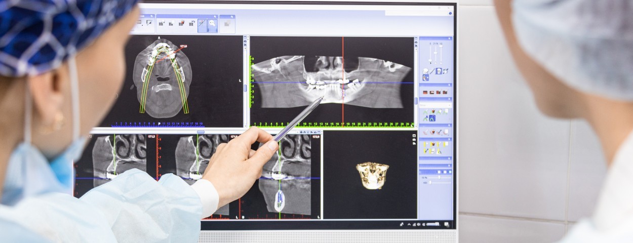
Cone Beam Computed Tomography (CBCT) provides 3D imaging of the facial skeleton and teeth. This section explains how CBCT captures images from multiple angles and creates a comprehensive view of the anatomical structures, ideal for diagnosing complex dental cases.
Patient Example: Incisive Canal
This section presents a case study demonstrating how CBCT can detect anomalies, such as a potential naso-palatine duct cyst, by providing detailed 3D views of the incisive canal and surrounding structures.
Artefacts: Noise and Blurring
CBCT images can suffer from noise and blurring caused by patient movement and X-ray scatter. This section explains how these artefacts affect image quality and offers techniques to reduce them, ensuring clearer diagnostic images.
Metal Artefacts
Metal restorations can create dark streaks and distortions in CBCT images due to beam hardening. This section covers methods to minimise the impact of metal artefacts and how to interpret images when such artefacts are present.
Spatial Resolution and Voxel Size
CBCT uses voxels to create 3D images, and smaller voxel sizes improve spatial resolution but require higher radiation doses. This section discusses the trade-off between image quality and patient safety, emphasising the use of the smallest acceptable resolution.
The Axial View
This section highlights the axial view, a horizontal slice through the imaged area, and its importance in evaluating structures such as the incisive canal and surrounding anatomy.
Justification
Every CBCT examination must be justified by ensuring that the benefits outweigh the risks. This section focuses on the criteria for radiological justification, including how CBCT can provide information that conventional radiographs cannot.
Patient Example: The ID Canal
This case study demonstrates how CBCT can be used to examine the inferior dental canal, helping with implant planning and avoiding nerve damage during surgery.
Sedentexct Basic Principles
This section outlines the Sedentexct principles, which provide guidelines for the safe and effective use of CBCT, focusing on radiation protection, justification, and optimisation.
Referral Guidelines
Referral criteria ensure that CBCT is only used when necessary. This section covers when to refer patients for CBCT imaging, emphasising the importance of considering lower-dose alternatives first.
Caries Detection
CBCT is not suitable for routine caries detection due to its limitations in showing the varying levels of decalcification. This section explains why conventional radiographs remain the preferred method for caries assessment.
Periodontics
In periodontics, CBCT can be useful for assessing bone defects, but it should not be used routinely for detecting bone levels. This section highlights when CBCT may be beneficial in managing complex periodontal cases.
Implantology
CBCT is commonly used in implantology for planning implant placement by providing accurate measurements of bone height, width, and proximity to anatomical structures such as the maxillary sinus and mandibular canal.
Endodontics
CBCT can help in identifying complex root canal systems and detecting periapical pathology when conventional radiographs are inconclusive. This section covers the indications for using CBCT in endodontic treatment planning.
Extractions
CBCT may be indicated for complex extractions, especially when there is a risk of damaging the inferior alveolar nerve during third molar surgery. This section discusses when CBCT is appropriate for extraction planning.
Other Criteria
CBCT can be used in other cases such as assessing dental trauma or skeletal abnormalities. This section covers additional scenarios where CBCT may provide very important diagnostic information.
Course Completion
Participants will complete a feedback survey, take a multiple-choice exam, and receive a GDC-compliant CPD certificate. The course emphasises the proper use of CBCT for safe and effective patient care.
You can copy and adapt this example PDP entry for your own needs and circumstances. The format complies with GDC guidance on PDP structure.
| PDP Learning or Maintenance need |
| IRMER training compliance |
| How does this relate to my field of practice? |
| I am involved in dental radiography/radiology and need to know about the uses of CBCT |
| Which development outcome(s) does it link to? |
| C D |
| What benefit will this have to my work? |
| Increase the range of options I can consider in radiography. |
| How will I meet this learning or maintenance need? |
| Take the Verified Learning IRMER CBCT course |
| When will I complete the activity? |